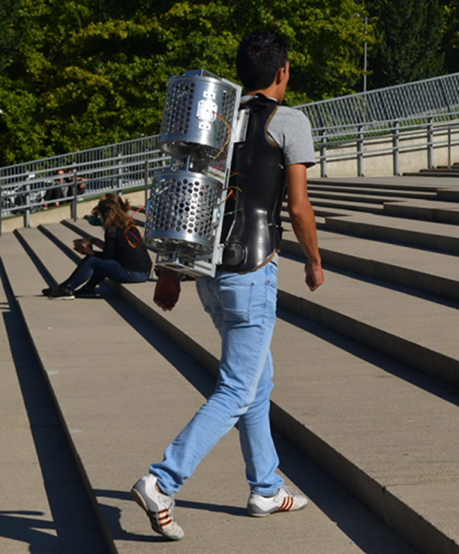
MRI scans can be made simpler and more affordable. Andrew Webb, Professor of MRI Physics at LUMC, is developing low-cost mobile MRI scanners that can be deployed in developing countries to detect hydrocephalus.
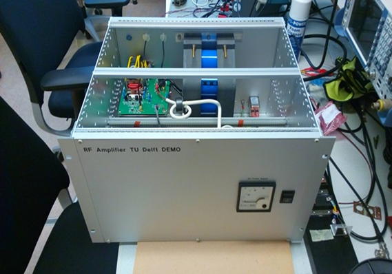
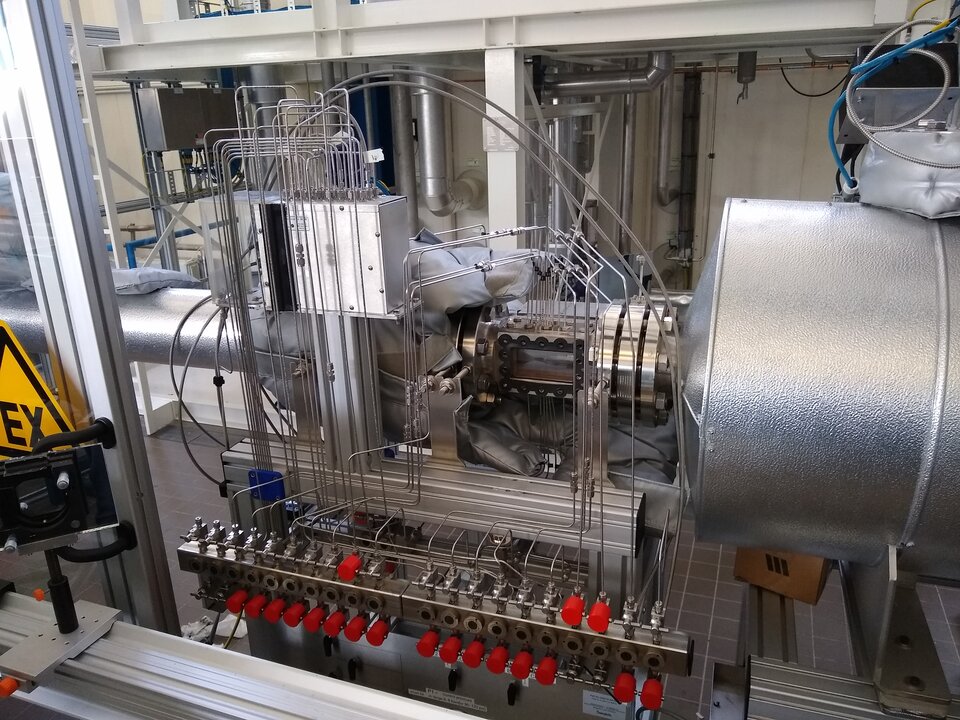
This project is a collaboration between Leiden and Delft. In Delft (Faculty of EEMCS), they are developing the necessary mathematics, and DEMO is working on, among other things, the RF Power Electronics – see photo below.
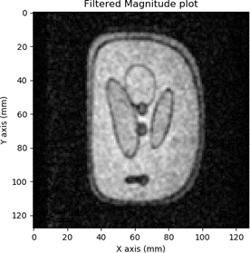
The first 3D images were produced using the setup at LUMC. For this, the RF amplifier previously developed was used, along with the new gradient amplifier. The first 'magnet' has been replaced by a new set of rings developed in Leiden, containing much smaller permanent magnets that together create the static field. This magnetic field is very uniform within the volume of the MRI, but to perform imaging, a gradient must be applied in the x, y, and z directions. During testing, the DEMO gradient amplifier was found to produce less noise than the commercial gradient amplifiers used, allowing for shorter scan times.
MRI scans could be better and less expensive. That is what Andrew Webb, an MRI Physics professor at Leiden University Medical Center, says. For his research, he recently received the 2017 Simon Stevin Master Award.
The largest technical-scientific research prize is worth half a million euros. Webb wants to use this to develop new MRI techniques to detect diseases such as Huntington's disease and Alzheimer's at a much earlier stage.
In addition, the professor develops inexpensive mobile MRI scanners that can be used in developing countries. For example, hospitals that do not normally have access to this type of medical equipment can still use a scanner that travels from place to place.
The inexpensive mobile MRI has a convenient side effect: it accelerates the process of the more expensive MRI scanners now used in hospitals. This relates to the software that Webb has to develop to control the mobile scanner. The best images are quickly filtered out of the device, which means that this could possibly accelerate the scanning process in the future.
DEMO
This project is a partnership with Leiden and EEMCS. Delft is developing the mathematics required to reconstruct the image.
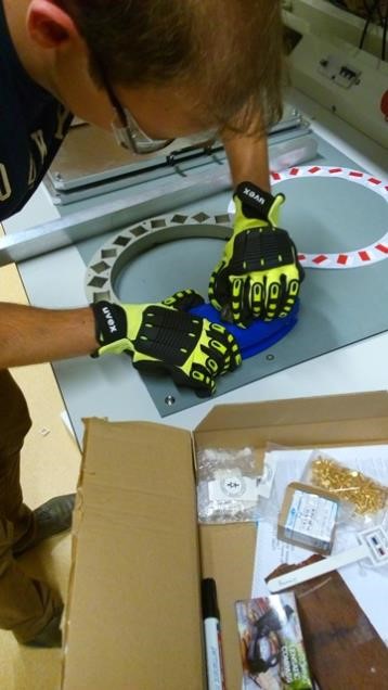
DEMO has designed the permanent magnet ring (see photo) and is also working on the RF power electronics (photo below). The project is still in the early stages of a 4-year research and we will soon be delivering the first parts. Improvements will then be made to the set-up based on the research results.
Update 2019
Since 2017, there have been many new developments in the low-cost MRI project. The first 3D images have now been made using the set-up at LUMC. This set-up uses the previously developed RF amplifier and the new gradient amplifier. The first ‘magnet’ has been replaced by a new set of rings developed in Leiden that contain a large number of far smaller permanent magnets that, together, create the static field.
This magnetic field is extremely steady within the volume of the MRI, but for imaging use, it is necessary to introduce a gradient in the x, y and z direction. In tests, the DEMO gradient amplifier was shown to produce less noise than the commercial gradient amplifiers used. This reduces the time required for a scan, and our amplifier is significantly cheaper even including the design costs.
The first magnet has returned to Delft and is now housed at the mathematics lab. A second MRI set-up with the same RF amplifier has now been built there. This set-up is used for research into a mathematical algorithm for making an MRI scan with a non-linear magnetic field. This makes it possible to use a magnetic field that is easier to make, resulting in an even cheaper system. The beauty of this new technique is that it can also be used on existing MRI scanners, to correct small variances in the magnetic field.
A Software Defined Radio (SDR) application has been created for the set-up in Delft. This generates the RF pulses that must be emitted and demodulates the received signal to low-frequency pulses, which consist of a real and imaginary part – exactly what the mathematicians need to create an image. The SDR was created using GNU Radio that generates Python code. Subsequently, an additional Python module was written, which can be used by researchers to carry out measurements.
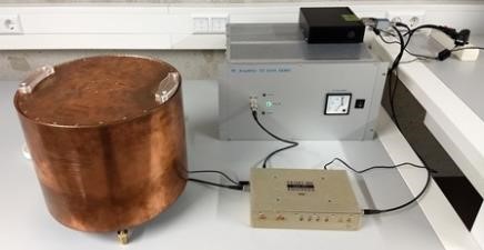
In order to measure the MRI signal, a signal is first emitted from the RF amplifier to a coil (in the copper cylinder). This signal consists of two RF pulses of 300 Vpp and 600 Vpp at 2.6 MHz. A bottle of water placed inside the coil is used as a test sample. The two pulses sent to the bottle cause the water itself to return a pulse shortly afterwards. This signal is received by the same coil and is so weak that it needs to be amplified more than 30,000x in order to use it. Suddenly we had a eureka moment. IT WORKS!!!!
Update 2020
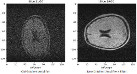
At the end of September, a huge step was taken in Leiden with the gradient amplifier for the MRI set-up. With an added filter, it now creates so little noise that its noise contribution in a scan is negligible. This was also true for DEMO’s RF power amplifier. This means that we are very close to the theoretical minimum noise. This is very important, because the signal strength in a Low Field MRI is very weak. Less noise means that a scan proceeds faster or that a higher resolution can be achieved in the same time frame.
The resolution required to scan hydrocephalus, for which it was designed, had already been achieved earlier.
Together with special coils wrapped onto a cylinder, the gradient amplifier allows a magnetic field to be created in the x, y, and z direction. This allows a point to be scanned in the patient to be determined in three dimensions. The set of points (“voxels”) realises the MRI scan.
Nice to know: in Berlin and at Penn State University (USA) the RF Power Amplifier is already being replicated to create a similar set-up there. The design has been released via open source so that inexpensive MRI scanners can be made worldwide.

