ERC Starting Grant for shedding light on neurodegenerative disease
Neurodegenerative diseases such as Huntington’s disease are incurable. These diseases are progressive brain disorders with severe effects on movement and mental functioning. How damage in the brain develops is far from understood however. Biophysicist Kristin Grußmayer has been awarded an ERC Starting Grant that enables her to search for causes by looking into brain cells and individual molecules.
Proteins clumping together
Neurodegenerative diseases such as Huntington’s disease are associated with a build-up of aberrant clumps of proteins – so-called aggregates – inside the brain. How damage in the brain develops is far from understood however, due to our lack of insight into early processes. Kristin Grußmayer expects to elucidate the role of protein aggregation and phase transitions, op te helderen, relevant for a broad variety of diseases
Recently, unusual liquid-liquid phase separation inside of brain cells has emerged as a possible cause for proteins clumping together in neurodegenerative disease. Grußmayer will investigate how different material states of aggregates form, using her expertise on studying individual molecules and taking fast 3D images of cells.

Lab experiments
Grußmayer will address the following key questions, with the use of cell models of Huntington’s disease along with well-controlled lab experiments: How do the physical and chemical properties of protein variants and their environment affect how they clump together and become harmful? What role does phase separation play in Huntington’s disease? How does spreading of proteins outside of cells interfere with protein assemblies in cells?
Deciphering Huntington’s disease
Huntington’s disease is caused by a gene defect resulting in aggregation-prone huntingtin protein. The diverse sizes and shapes of the aggregates pose a remarkable challenge, and multiple complementary approaches are needed to unravel their shape, structure and characteristics. Grußmayer will develop multimodal AI-informed quantitative microscopy and nanoscopy to decipher how aggregates in neurodegenerative disease form and mature. This will allow her to connect what is going on at the molecular level to the disease symptoms at the cell level. Importantly, her group will work without directly attached dyes and exploit virtually stained quantitative microscopy, interfering with aggregation processes as little as possible.

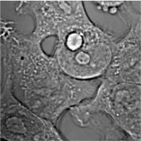
Header image credits: KATERYNA KON/SCIENCE PHOTO LIBRARY/GETTY IMAGES

